Support for the practice of following moles with pictures was published in the Journal of Pediatrics.
Spitz nevi, moles that needs to be differentiated from melanoma, were followed with clinical images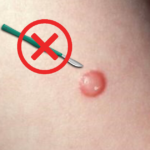 and dermoscopy. The conclusion was that particularly with younger children, typical Spitz nevi can be followed with sequential images and avoid surgery.
The following editorial was published in Practice Update:
Evolution of Spitz Nevi:
Nazan Emiroglu , M.D.,* Pelin Yıldız, M.D.,† Dilek Biyik Ozkaya, M.D.,* Anıl Gulsel
Bahalı, M.D.,* Ozlem Su, M.D.,* and Nahide Onsun, M.D.* Pediatric Dermatology 1–8, 2017
Editorial
Gary D. Lichten, M.D.
Seventy Spitz nevi were studied: 43 initially excised, 26 involuted, and 6 remained.
Twenty-seven were followed clinically and with dermoscopy images. The dermoscopy pattern, most frequently observed, was globular, followed by starburst. The age group was younger with the Spitz nevi that involuted: age14.3 (range 4.6 years), contrasted with mean age of the stable group was 19.0 (range 4.2 years). The range for involution of lesions was 21-27 months.
Although the number of cases were limited, the practice of following Spitz nevi in younger children with clinical and with dermoscopy images is laudable. When viewing a suspicious pigmented lesion, because of the fear of missing a melanoma, we choose surgical removal as the first option. When given a choice, patients and parents frequently opt to be followed with photographs, and thus avoid a procedure and scar. With typical Spitz nevi in children as well as atypical nevi, I frequently follow nevi with dermoscopy images.
Although the authors followed Spitz nevi every 6 months, for atypical nevi, I use short-term, three month, monitoring. Patients are engaged with taking and comparing images monthly with an application, with instructions to return immediately for excision if any change is noted. Flat, atypical nevi, excluding those with any melanoma specific features ( atypical or negative network, irregular globes or streaks, blue-white colors, regression structures) are photographed with dermoscopy initially and again at 3 months. I recommend the free application, CompariSkinFree,
CompariSkin, which enables patients to take consistent images and compare for changes with side-by-side and overlaid images within one minute.
Sequential dermoscopy images tell a story of change, which can be reassuring to the patient and parents for Spitz nevi, as well as atypical pigmented nevi.
|
|
Gary Lichten, M.D.
Pingback: http://vioglichfu.7m.pl/